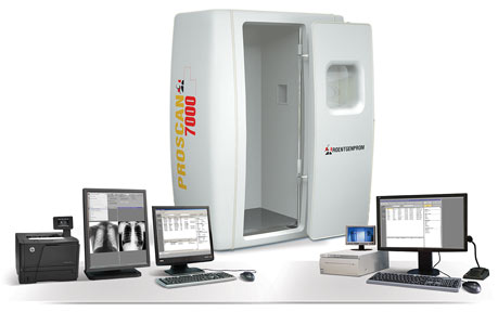
Operating principle: To register the X-ray radiation, which passed through the chest, X-ray machine ProScan uses a silicon linear detector. To make a fluorography, the detector is being moved along the chest horizontally together with a sector-shaped X-ray formed by the slit collimator. High quality images, lowest dose of investigation, convenient conditions for patients and personnel, easy requirements to X-ray room, all these parameters make this chest X-ray machine to be better solution for mass screening compare to bucky based solutions.
Specific features of fluorography units ProScan®-2000 and ProScan®-7000
Radiologist and assistant workstations. Both stations are equipped with the latest models of computers. Assistant workstation is equipped with a professional graphic monitor 21 inches, and that of the radiologist with the monitor 23 inches and a graphic one 21 inches. On the enquiry of a customer both versions of ProScanâ can be equipped with professional medical monochrome monitor not less than 20 inches.
The ProScan software enables the doctor to examine the images after fluorography on the big monitor, at the same time using the second one for work with the database. A professional graphical printer gives 256 gray shades and high spatial resolution on thermal paper. Herewith the image is equally informative on the monitor as well as on a hard copy. To print out periodical reports, the device is equipped with a laser office printer. The unit has special external storage element on hard disks for long-term storing of images. Capacity of storage device allows place on hard disks image archives accumulated within 10 years of unit operation. Built-in storage software provides particular reliability of saved information and absolute archive reliability in case if one of hard disks fails.
ProScan® Software meets the requirements of the DICOM 3.0. The program is being developed in close cooperation with radiologists that is why it contains not only generally accepted formalized protocols, but also the necessary forms of periodical reports. The program offers wide possibilities for processing of obtained images through special filters to detect tuberculosis.